Browsing Tag
imaging
6 posts
Imaging the adolescent heart provides ‘normal’ reference values for clinical practice
Cardiac magnetic resonance images used to study distinct aspects of cardiac anatomy and function. (A and B) Atria. (B and C) Ventricles. (E and F) Cardiac tissue characterization. Credit: CNIC Magnetic resonance imaging (MRI) has allowed scientists at the Centro Nacional de Investigaciones Cardiovasculares (CNIC) to produce an accurate picture of the healthy heart in
March 3, 2023
Imaging the adolescent heart
Cardiovascular magnetic resonance (CMR) is an accurate method for determining the heart’s architecture, function, and tissue makeup. There aren’t many studies that provide CMR reference levels for adolescents, though. A new study by the scientists at the Centro Nacional de Investigaciones Cardiovasculares (CNIC), aims to provide sex-specific CMR reference values for biventricular and atrial dimensions
March 3, 2023
Real-time imaging of Arc/Arg3.1 transcription ex vivo reveals input-specific immediate early gene dynamics
Checking if the site connection is secure www.pnas.org needs to review the security of your connection before proceeding.
September 12, 2022
Imaging Pinpoints Markers of Anxiety Related to Parkinson’s
The insula and frontal cortex are involved in the development of anxiety in adults with Parkinson’s disease, according to imaging data from 108 individuals. Anxiety occurs in approximately 31% of Parkinson’s disease patients, but the underlying mechanisms are not well understood, wrote Nacim Betrouni, MD, of the University of Lille, France, and colleagues. Previous research…
February 20, 2022
EO Vista imaging sensor for Space Force weather satellites passes design review
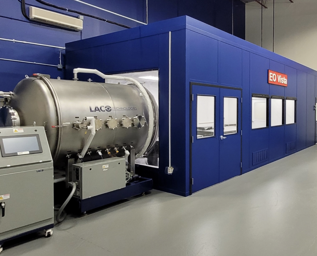
by Sandra Erwin — January 31, 2022 EO Vista’s electro-optical infrared payload integration, test and calibration Facility. Credit: EO Vista EO Vista is supplying sensors to General Atomics Electromagnetic Systems, one of three contractors competing in the U.S. Space Force Electro-Optical Infrared Weather System program WASHINGTON — An imaging sensor developed by EO Vista for…
January 31, 2022
Optical imaging highlights metabolic interactions that make pancreatic tumor cells grow
Representative NAD(P)H fluorescence intensity images of pancreatic cancer cells cultured as 3D organoids (shown in magenta) and PSC non-cancer cells (shown in cyan) from cocultures obtained on days 1 through 4 (top). The corresponding optical redox ratio from the same images is also shown (bottom). Credit: Morgridge Institute for Research Pancreatic cancer is a rare,…
January 21, 2022


