


Making an embryo is much like making a Lego castle: In the same way that a castle needs turrets and gargoyles and a moat, you need two legs and two eyes and a heart.
Except unlike the Lego Camelot, you don’t come with a picture on the box of what you’re meant to look like, much less an instruction manual—and you’re not going to be the one to assemble the structure. Instead, you’ll sit back and wait for the Lego pieces to organize themselves. Our cells, our little Lego pieces, assembled themselves. What’s even more astonishing is that when they get it right, all those cells get it right in broadly the same ways: We all managed to come out with the characteristic shape and proportions appropriate to our species (we can all spot a regulation-issue chicken, frog, mouse, or human shape).
There’s no gene for “two arms and two legs.”
So how did all our initial progenitor cells know how to organize into us—forming our eyeballs, legs, fingers and all that in the right place and the right order? Who gave them the blueprints to check that all those fingers or fins or beaks weren’t too giant, or too tiny, or of wildly different lengths? Most important—how did they know when to stop?
Well, you might be thinking: That’s what DNA is for. Not true. You can search all the As, Ts, Cs, and Gs in your genome, turn ’em upside down and shake the spare change out of their pockets—you won’t find the instructions for anatomy.1 You’ll find plenty of specs—code that tells you about the color of hair a baby will have, its skin, its eyes. You’ll find nothing about how many eyes. There’s no gene for two eyeballs. There’s no gene for “the eyeballs need to go on the front of the head.” There’s also no gene for “two arms and two legs, kind of this far apart, feet in the front and butt in the back.” It’s simply not possible to recover the shape of an organism solely from reading a printout of its genome.
So if not genes, what does control your shape?
The question had begun to assemble itself in Mike Levin’s head when he was still a child, wondering how an entire person could be assembled from an egg. As it happened, Levin needed a topic for his Ph.D. thesis at Harvard Medical School, and in the early 1990s, there was still one aspect of how human beings are shaped in the womb that remained a total mystery: how embryos tell left from right. There had been theories, but never a smoking gun.
For a grad student, this was tempting, low-hanging fruit. So Levin, now a developmental and synthetic biologist at Tufts University, began to investigate how all those cells, without having brains, nonetheless seemed to know their left from their right. And make no mistake—their ability to distinguish left from right during development is critical to our survival. From the outside we may pull off the illusion of symmetry—two eyes, two ears, two arms, and two legs, same on one side as we are on the other. But inside, it’s a different story. You probably know that your heart and stomach lean left, and that your right side houses your liver, appendix, and pancreas. For about 1 in 20,000 people, this whole image is flipped.2 And that’s absolutely fine! They don’t usually have health issues (apart from overzealous researchers poking and prodding them to understand their condition, known as situs inversus). However, when just some of the bits are flipped, you have a problem. Jumbling up the body’s precise internal asymmetry, particularly when it affects the fastidious plumbing of the heart, is the origin of many congenital heart defects and other life-threatening syndromes.
Understanding what caused any of this—the correct pattern, the flipped pattern, the jumbled pattern—was a longstanding, perennially interesting unsolved mystery. Why does the heart go on the left and not the right? And how does the body know to develop this way? No one had been able to finger a specific molecular component, so there was no hint of a genetic cause. Genes couldn’t be the whole story, anyway. After all, genetic information is not spatial. The genome can’t tell left from right. It seemed to Levin, from studying the old ion current papers, that electricity was somehow fundamental to establishing the polarity of a cell.3 But how?
Decades of work had cataloged every ion that shuttled in and out of developing embryos of every species and identified the ion channels that send them through the zygote and the blastomeres it calves off into as it begins to diversify into the developing embryo. A funny thing happens to the ions and ion channels in the cell during this transition: They all mysteriously change. Some pop into existence, others disappear then reappear, and their currents wax and wane with those disappearing and reappearing acts.
Another clue to the functional importance of these weird ion events was what happened when you interfered with them. Disrupting even seemingly minor sodium currents resulted in a “rosette,” which is an abnormal embryo that “appears to have lost its spatial orientation,” Italian biologist Elisabetta Tosti found in her own research. She concluded that the currents during and after fertilization are crucial for correct embryo development.4 Messing with potassium currents also led to developmental defects, further evidence that ion movements are crucial for an embryo. But no one had been able to assemble this jumble of interesting pieces into a coherent whole.
By the time the 21st century got underway, Levin could ask these questions in his own lab at the Forsyth Institute, at Harvard. How was electricity setting the polarity of a cell? He and Ken Robinson, working at Purdue University, discovered a proton pump. Protons are hydrogen ions. This pump specializes in making sure hydrogen and potassium are kept in strict ratios. On an unfertilized frog egg, proton pumps are speckled evenly around the whole surface.
Genetic information is not spatial. The genome can’t tell left from right.
But when Levin and Robinson checked on these pumps after fertilization, they found something strange: All the channels had started drifting over to one side of the egg, where they smashed themselves into a tight little clique. No one had ever seen anything like this. When the pumps gathered on one side of the egg, it meant hydrogen ions were only able to get in or out of the cell on that one side. This created a voltage, and it happened really soon after fertilization, when the frog embryo consisted of only four cells. Could this be the answer they were looking for?
When scientists think they have found a causative agent like this, their next step is to try to figure out an experiment that can disprove their idea. Levin and Robinson decided to see what would happen if they prevented the proton pumps from drifting out of perfect symmetry after fertilization. To do that, they added extra proton pumps or potassium channels to the developing embryo to even out their distribution, mimicking the smooth distribution on an egg. If the researchers were right, this evenness would wreak havoc on the embryo’s ability to discern left from right. And they were correct. The embryos with the extra proton pumps were all messed up, as likely to have their hearts on the right as the left. The proton pump was clearly essential to kicking off the difference between left and right sides.5
But it was also changing the membrane voltage. That was weird. A change in membrane potential is how nerves send action potentials. But why was a brand-new embryo changing membrane potentials? What possible use could that be, when it hadn’t even developed nerves yet? Levin wondered if this voltage was part of the system an embryo used to tell its constituent cells to become different kinds of tissue. This idea is articulated by the biologist Mina Bissel: If all our cells have the same exact genes in them, then why do some of them do one thing when others do other things? How do some of them become bone cells and others skin, or nerves?
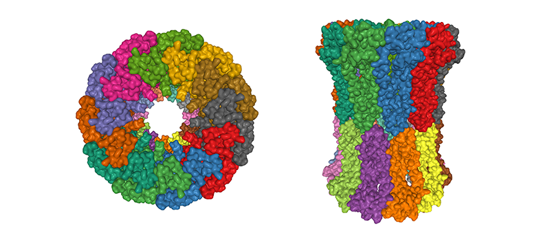
Gap junctions are an unusual kind of ion channel. They start to form the moment the fusion of egg and sperm creates the first joint cell, known as a zygote. Right away, the gap junctions establish a cellular intranet quite unrelated to the nervous system (which in a zygote of course does not yet exist). Unlike other ion channels which let in and keep out various molecules, gap junctions simply create direct connections cell to cell to cell.6 They poke between neighboring cells, creating a sneaky door only they share, a bit like adjoining hotel rooms.
That means that as cells differentiate, each new cell that cleaves off is already connected to the cells around it. Long before nerve cells develop synapses, our non-excitable embryonic cells are equipped with this other, much faster, more electrical way of communicating.
Levin had long suspected that these gap junctions were involved in how an organism decides how to shape itself. In his earliest days of investigating left-right patterning, he had found that turning off gap junctions also messed up normal asymmetry. Later, he and a postdoc, Taisaku Nogi, found gap junctions were the culprit in the unparalleled regenerative superpowers of a weird little sea worm called a planarian.7 This small flat worm can regrow no matter how fine you chop it—and it only takes about a week to return to fully functional normalcy. Nogi and Levin realized that gap junctions could explain how this re-patterning information could spread so quickly over thousands of cells.
So that was two different animals, then, in which the gap junctions seemed to enable long-distance messages without a nervous system. In some ways, this system was actually better than a nervous system. When two cells are connected in this way, each cell has direct, privileged access to the other’s internal informational universe. What one cell knows or experiences diffuses immediately through the connecting door for its neighbor to know or experience too. The effect is close to telepathy.
This was when Levin first began to formulate his theory of the bioelectric code. The membrane voltage carried information, and the gap junctions formed the body-wide network—the electrical network that was not the nervous system—that sent that information around the body. It wasn’t much of a stretch to think of this information as taking the form of a code.
That code controls the complicated biological processes that formed you in the womb, by executing a controlled program of cell growth and death. It is the reason you retain that same shape throughout your entire life; it prunes your dividing cells so you keep being recognisably you. It wasn’t the only important thing—biomechanics, biochemicals, and all the rest mattered too. But just as the neural code was mooted to govern behavior and perception, and the genetic code governs heritable traits, the bioelectric code was how the body told itself about its form. 
Sally Adee is an independent science and technology journalist based in London. Her first book, We Are Electric is out now in the U.S. and the U.K.
Adapted from We Are Electric: Inside the 200-Year Hunt for Our Body’s Bioelectric Code, and What the Future Holds by Sally Adee. Copyright © 2023. Available from Hachette Books, an imprint of Hachette Book Group, Inc.
Lead image: Kravpak / Shutterstock
References
1. Kwok, R. The shape of life: Biology’s biggest mystery. New Scientist 21538-41 (2012).
2. Zimmer, C. Growing left, growing right. The New York Times (2013).
3. Nuccitelli, R. Ionic currents in development. New York: International Society of Developmental Bi ologists (1986).
4. Tosti, E., Boni, R., & Gallo, A. Ion currents in embryo development. Birth Defects Research Part C 1086–18 (2016).
5. Levin, M., Thorlin, T., Robinson, K.R., Nogi, T., & Mercola, M. Asymmetries in H+/K+-ATPase and cell membrane potentials comprise a very early step in left-right patterning. Cell 11277-89 (2002).
6. Moody, W.J., Simoncini, L., Coombs, J.L., Spruce, A.E., & Villaz, M. Development of ion channels in early embryos. Journal of Neurobiology 22674-684 (1991).
7. Nogi, T. & Levin, M. Characterization of innexin gene expression and functional roles of gap-junctional communication in planarian regeneration. Developmental Biology 287314-335 (2005).
![]()
![]()
Get the Nautilus newsletter
The newest and most popular articles delivered right to your inbox!
Note: This article have been indexed to our site. We do not claim legitimacy, ownership or copyright of any of the content above. To see the article at original source Click Here

Beef Tallow For Skin - Whipped Tallow Balm with Organic Jojoba Oil (Unscented/Herb-Infused), Grass Fed Beef Tallow Face Moisturizer for Eczema, Baby, Lip Balm - Lotion For Extremely Dry Skin
$29.99 (as of March 14, 2025 20:23 GMT +00:00 - More infoProduct prices and availability are accurate as of the date/time indicated and are subject to change. Any price and availability information displayed on [relevant Amazon Site(s), as applicable] at the time of purchase will apply to the purchase of this product.)
Pet Steam Brush for Dog & Cat – 3-in-1 Spray Hair Removal Comb – Steam Brush for Shedding & Grooming – Water Brush for Long & Short Haired Pets – Spritz Defur Comb – Includes Waterless Shampoo
$24.99 (as of March 14, 2025 19:47 GMT +00:00 - More infoProduct prices and availability are accurate as of the date/time indicated and are subject to change. Any price and availability information displayed on [relevant Amazon Site(s), as applicable] at the time of purchase will apply to the purchase of this product.)
VYG Bunny - Realistic Bunny Toy, Bunby - My Realistic Bunny Toy for Kids (Brown)
$7.25 (as of March 14, 2025 19:47 GMT +00:00 - More infoProduct prices and availability are accurate as of the date/time indicated and are subject to change. Any price and availability information displayed on [relevant Amazon Site(s), as applicable] at the time of purchase will apply to the purchase of this product.)
Abardsion Women's Casual Basic Going Out Crop Tops Slim Fit Short Sleeve Crew Neck Tight T Shirts
$9.97 (as of March 14, 2025 20:23 GMT +00:00 - More infoProduct prices and availability are accurate as of the date/time indicated and are subject to change. Any price and availability information displayed on [relevant Amazon Site(s), as applicable] at the time of purchase will apply to the purchase of this product.)